Stabilization of odontoid and axis (C2) fractures
An odontoid fracture is the most common break of the axis (C2). It happens after a fall in an older adult, a road accident or a whiplash injury. The cord runs millimetres from the fracture line, so prompt assessment matters. Not all of them need surgery. Some heal with a rigid cervical collar for several weeks. Others are stabilised with an anterior screw through the odontoid or with posterior C1-C2 fixation (Goel-Harms technique). Fracture type, age, bone quality and CT and MRI findings drive the decision. In the Alicante and Benidorm clinics each case is reviewed individually. Step-by-step details are below.
What is an odontoid or axis (C2) fracture?
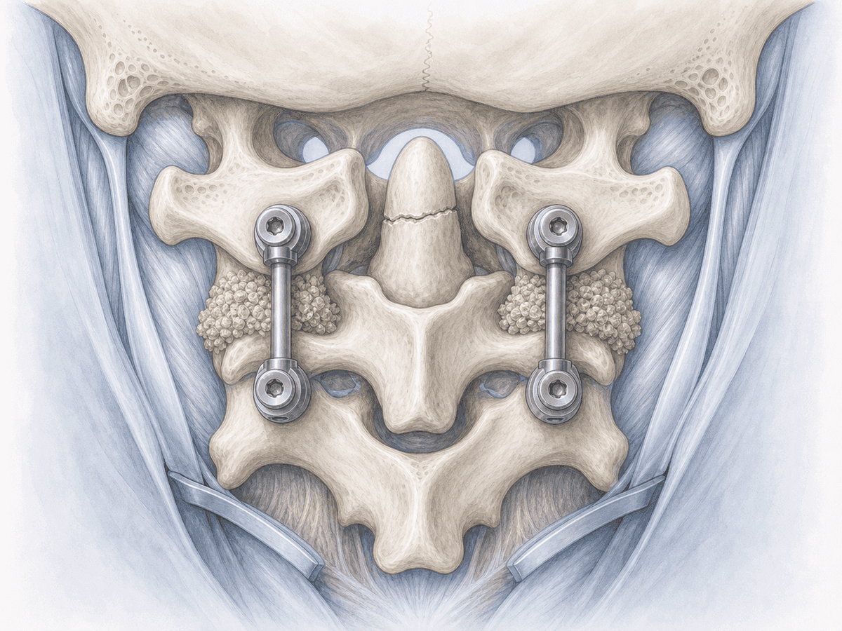
The odontoid (or dens) is a small bony projection of the second cervical vertebra, also called the axis or C2. It fits into the first vertebra (atlas, C1) and allows the head to rotate from side to side. A fall onto the face or forehead, a whiplash injury or a blow to the neck can break it.
The most widely used classification is Anderson and D'Alonzo's, based on the CT scan, and it guides treatment. Type I is a fracture at the tip of the dens: it is uncommon and almost always stable, so it is usually managed with a collar. Type II is a fracture at the neck of the dens: it is the most frequent (60–80 %), the hardest to heal and the one most often operated on, especially if displaced. Type III enters the C2 body: the bony contact area is large and it usually heals well with a collar over 6 to 12 weeks.
There is also the hangman's fracture (traumatic spondylolisthesis of the axis), which involves the C2 pars. Today it is seen mostly after road accidents and falls. The Levine and Edwards classification distinguishes four types: stable ones (I and many II) are usually managed with a collar, while unstable patterns (IIA and III) require surgery.
Stabilisation aims at three things: realigning the bone, keeping it still while it heals and protecting the nearby spinal cord. As with occipitocervical fusion, the technique varies according to the fracture pattern. To review your case you can request an assessment with Dr. Ben Ghezala.
How it presents after a fall or accident
After a fall, a road accident or a whiplash injury, the typical signs are:
When does an odontoid or axis fracture need surgery?
Many fractures are managed with a rigid collar for 6 to 12 weeks. In older adults, the halo vest is now rarely chosen: it increases respiratory complications and mortality. Surgery is considered when there is:
How is the surgery performed?
1.Before surgery
The patient usually arrives with a rigid collar after the emergency department. Imaging is completed with high-resolution cervical CT, MRI to rule out transverse ligament or cord injury, and CT angiography if posterior fixation is planned, to map the vertebral arteries.
We assess the fracture pattern, bone quality (key in osteoporosis) and overall patient condition. Options, benefits and risks are explained to patient and family. Anaesthesia adjusts chronic medication and reviews comorbidities before consent.
2.During surgery
Surgery is performed under general anaesthesia, almost always with neuromonitoring to track cord and root function.
The anterior odontoid screw is placed through a small incision at the front of the neck. Under fluoroscopic guidance a cannulated screw is advanced across the fracture and anchored at the tip of the dens. It preserves C1-C2 rotation and is reserved for favourable fracture patterns and good-quality bone.
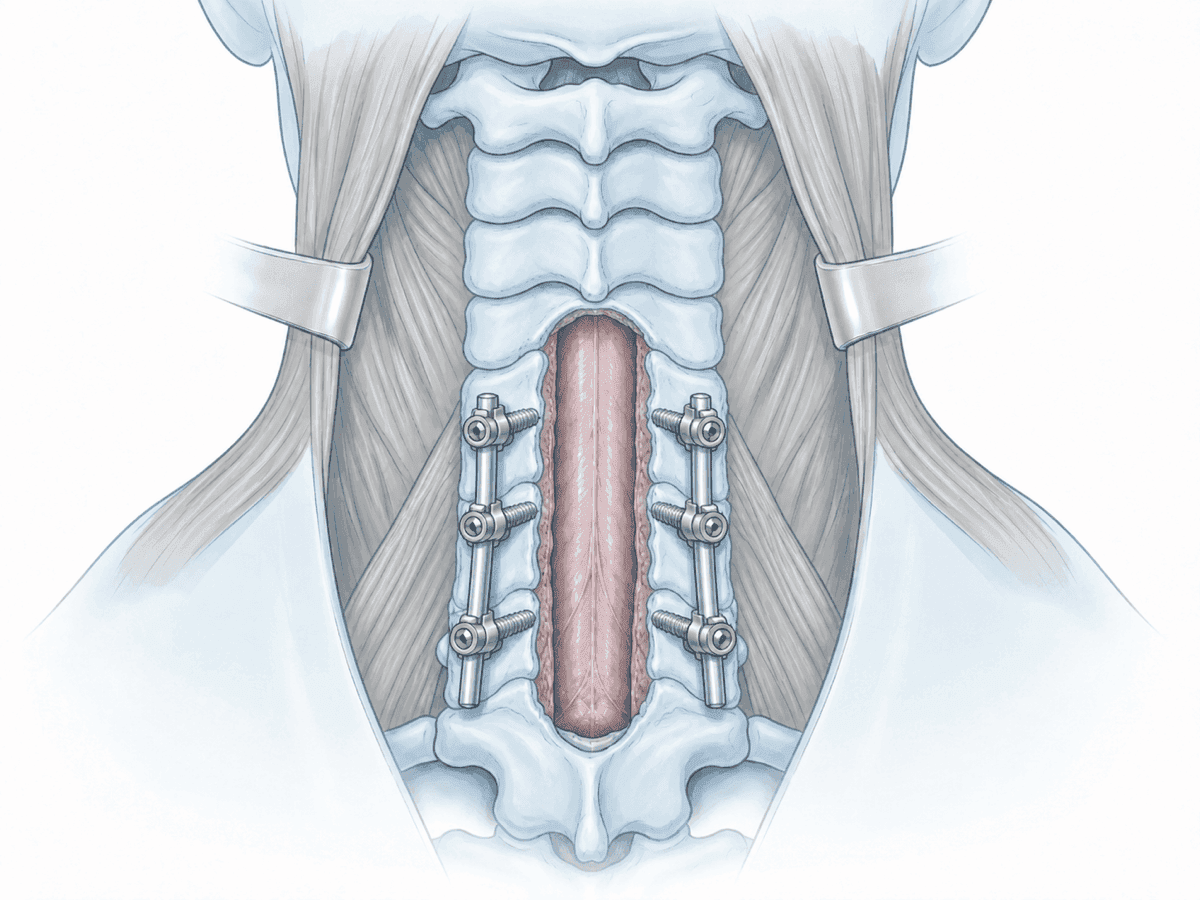
Posterior C1-C2 fixation (Goel-Harms) is performed prone. We place screws in the C1 lateral masses and C2 pedicles, connected by two titanium rods. Posterolateral bone graft is added. It is the most stable technique, preferred in osteoporotic bone, transverse ligament injury and in most older patients. The Magerl technique, with transarticular screws, is an alternative when vascular anatomy allows.
For hangman's fractures, depending on the pattern, C2 isthmus screw fixation (motion-preserving) is enough, or a short C2-C3 fusion is performed from the front or the back.
3.Immediately after
After surgery the patient stays in recovery. Vital signs, neurological exam, airway and pain control are monitored. Neck discomfort and, in anterior approaches, swallowing irritation are normal.
Mobilisation usually starts the same day or the next, assisted and with a soft or semi-rigid collar for several weeks. A postoperative CT confirms screw position before discharge. Hospital stay is typically 3 to 7 days depending on age, comorbidities and progress.
Recovery, follow-up and return to normal life
Healing of a C2 fracture takes time. In the first weeks a collar is used and sudden neck rotation, loads and sports are avoided. Clinical and CT follow-up at 6 weeks, 3 months and 6 months confirm that the bone is healing.
Return to office-based work is usually considered between 4 and 8 weeks if pain is controlled and imaging is favourable. Physically demanding jobs, contact sports and professional driving may need 3 to 6 months.
Physiotherapy is introduced gradually. We start with gentle isometric exercises and progress to mobility and strength, avoiding extreme rotation until cleared. Fever, marked wound redness, disproportionate pain, swallowing difficulty or new weakness call for prompt review.
Risks and possible complications
As with any surgery, there are risks. General ones include anaesthesia-related issues, wound infection, bleeding, haematoma and venous thrombosis.
Specific risks include nerve root injury or, rarely, spinal cord injury; partial symptom persistence; non-union (pseudoarthrosis), screw malposition or loosening, especially in osteoporotic bone.
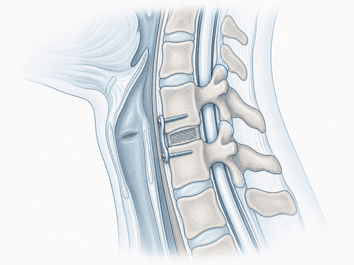
The anterior approach may cause transient swallowing discomfort, injury to oesophagus, trachea, cervical vessels or recurrent nerve. In the posterior approach the most serious risk is vertebral artery injury; residual neck pain and loss of about half of cervical rotation after C1-C2 fusion are also possible.
In elderly or frail patients, medical complications (respiratory, cardiovascular, delirium) are more likely. The decision to operate is always weighed with patient and family.
Frequently asked questions
Other related treatments

Do these symptoms sound familiar?
If you recognise yourself in some of these symptoms and your pain is starting to limit your daily life, we can review your case in a personalised consultation. Dr. Ben Ghezala will assess your clinical history and imaging studies to help you decide the best treatment option for you.
Request a consultation with Dr. Ben Ghezala