Revision of failed cervical fusion (cervical pseudarthrosis)
Revision of a failed cervical fusion is indicated when previous cervical surgery, usually an ACDF, has not achieved bony fusion (pseudarthrosis) and pain persists, hardware loosens or new neurological symptoms appear. We understand how disheartening it is when surgery has not solved the pain and how unsettling it feels to consider a second operation. Our role is to offer a careful second-opinion assessment, reviewing your previous reports, current imaging and clinical course. This guide summarises when revision should be considered, the available technical options and what to expect from the cervical salvage process.
What is the revision of a failed cervical fusion?
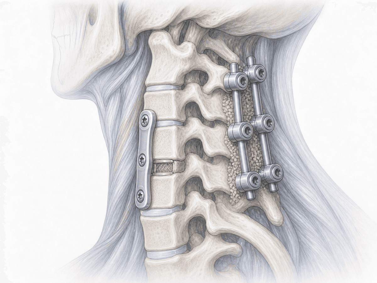
Revision of a failed cervical fusion is a salvage operation aimed at treating a previous cervical fusion that has not consolidated. When bone fails to bridge the operated segment the condition is called pseudarthrosis. The treated level remains abnormally mobile, pain returns or never fully resolves and, in some cases, the screws or interbody cage become loose.
This is a familiar scenario for our team. Dr. Ben Ghezala has authored dedicated chapters on pseudarthrosis in reference German textbooks such as Spinale Neurochirurgie (Schattauer, 2009) and Wirbelsäule Interdisziplinär (Schattauer/Thieme, 2017), which guides planning when a first surgery has not met its goals.
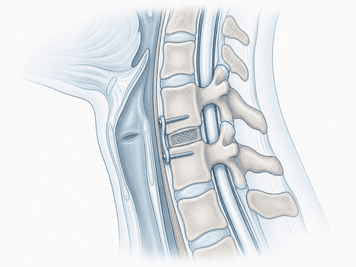
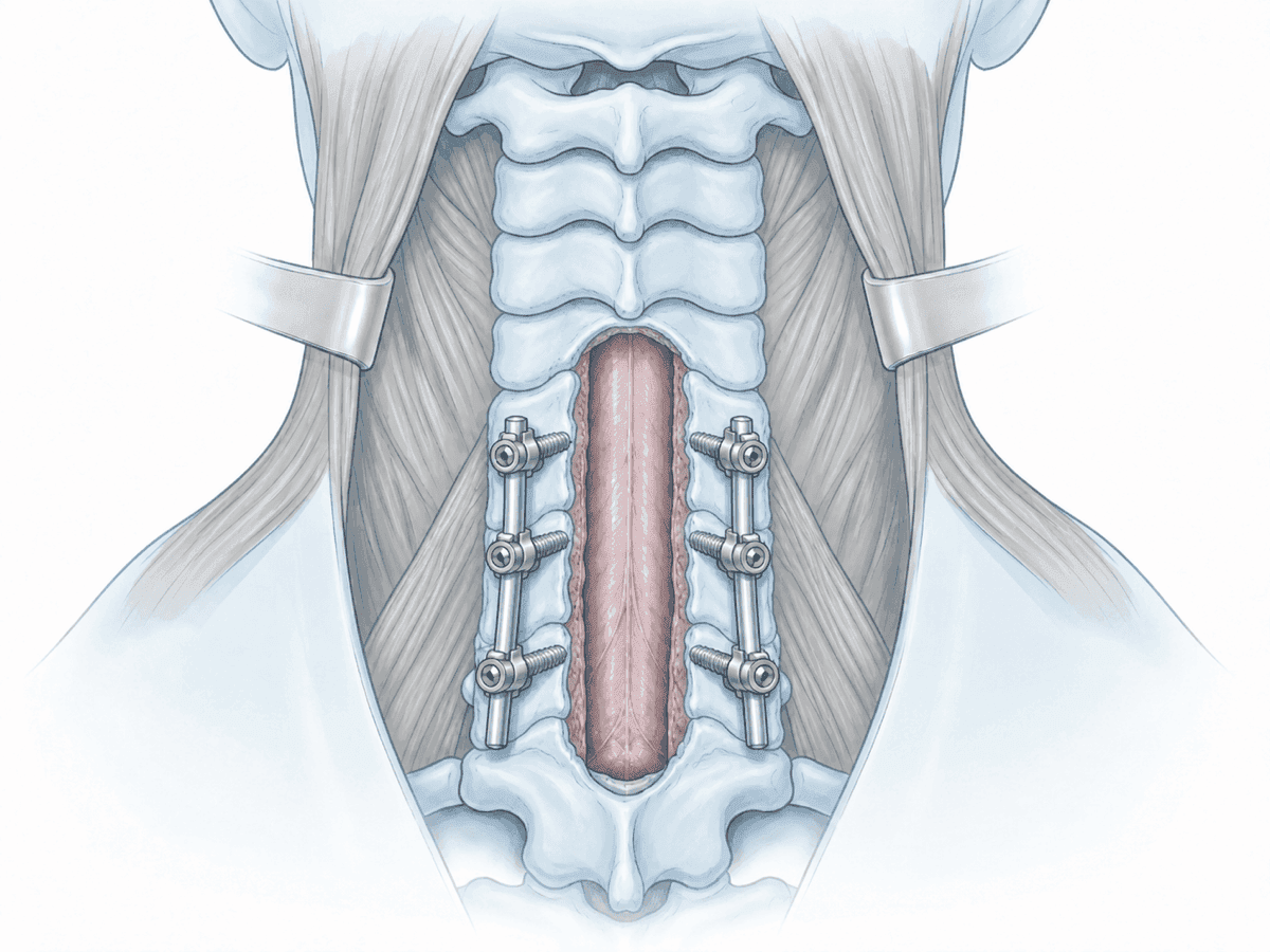
The strategy depends on each case. In some patients the answer is to reinforce the fusion through a posterior approach, with posterior cervical fusion and lateral mass or pedicle screw instrumentation. In others, it is preferable to re-operate from the front, revising the previous anterior cervical discectomy and fusion (ACDF), removing the plate, extending the decompression and placing a new graft. When poor bone quality, multiple levels or kyphosis coexist, combined anterior plus posterior approaches are considered.
To review your case, you can request a second opinion with Dr. Ben Ghezala.
Symptoms and signs of a failed cervical fusion
After a cervical fusion, certain signs should raise suspicion of pseudarthrosis or hardware failure:
When is revision surgery indicated?
How is the procedure performed?
1.Preoperative preparation
Preparation includes a complete review of the previous surgery: operative report, implant type, recovery and prior tests. Fine-cut cervical CT, MRI and flexion-extension X-rays are usually requested. Bone quality, hardware status and the presence of kyphosis are assessed. Medication is adjusted, postoperative analgesia is planned and the chosen strategy and alternatives are explained in detail.
2.During the procedure
The procedure is performed under general anaesthesia with neurophysiological monitoring. If a posterior salvage is chosen, the back of the neck is exposed, lateral masses and pedicles are identified and screws and rods are placed to stabilise the segment; bone graft is usually added and, in selected cases, biologics are used to promote fusion.
In an anterior revision, the previous plate and implant are carefully removed, decompression is completed and a new graft or cage is placed, with an updated plate. When kyphosis or several affected levels coexist, a combined anterior and posterior approach in one or two surgical sessions may be considered depending on the case.
3.Immediate postoperative period
After surgery the patient spends a short time in the recovery area before returning to the ward. Pain is controlled, a postoperative X-ray or CT is obtained and progressive mobilisation begins, usually with a soft collar during the first weeks. Hospital stay is typically 2–4 days, slightly longer than in primary surgery, depending on the complexity of the salvage and the clinical course.
Recovery after cervical revision surgery
Recovery after a revision is somewhat slower than after primary surgery. Daily activities are gradually resumed within a few days. Office work usually restarts between 4 and 6 weeks; physical jobs require more time and an individual assessment.
Bony fusion takes several months to consolidate. Heavy strain, impact sports and prolonged postures are avoided during the first three months. Guided rehabilitation, good ergonomics and clinical-radiological follow-up are key to achieving a stable fusion. Fever, severe pain, breathing difficulty or new neurological symptoms warrant prompt medical review.
Risks and possible complications
Any surgery carries general risks such as infection, bleeding, thrombosis or anaesthesia-related complications. Cervical revision surgery adds extra risk due to scar tissue, the presence of previous implants and the anatomy modified by the first operation.
Specific risks include nerve root or spinal cord injury, oesophageal or tracheal injury in anterior revisions (uncommon but more likely than in primary surgery), temporary swallowing difficulty or voice changes, vascular injury, persistent pseudarthrosis requiring further salvage and accelerated adjacent-segment degeneration. Each patient receives an individualised assessment to minimise these risks.
Frequently asked questions
Other related treatments

Do these symptoms sound familiar?
If you recognise yourself in some of these symptoms and your pain is starting to limit your daily life, we can review your case in a personalised consultation. Dr. Ben Ghezala will assess your clinical history and imaging studies to help you decide the best treatment option for you.
Request a consultation with Dr. Ben Ghezala